- 01: Introduction
- 02: History
- 03: Propellants, Firearms, and Ammunition Development
- 04: Modern Firearms Manufacture
- 05: Small Arms Ammunition
- 06: Evidence Handling Procedures
- 07: Equipment and Instrumentation
- Introduction
- Objectives
- AFTE Knowledge and Ability Factors
- Measurement, Standards, and Accuracy
- Weight, Force, and Their Measurement
- Dimensional Data and Measurement Tools
- Muzzle Velocity of Bullets
- Stereo and Comparison Microscopes
- Small Tools and Supplies
- Imaging Equipment
- Field Support Equipment
- Computer-Based Technologies
- Firing Facilities
- Selected Bibliography
- 08: Examination of Firearms
- 09: Cartridge and Shotshell Examination
- 10: Characterization and Evaluation of Fired Projectiles
- 11: Bullet Comparison and Identification
- 12: Gunshot Residue and Distance Determination
- 13: Toolmark Identification
- 14: Communicating Results
- Resources


Comparison Microscopy
Home > Equipment and Instrumentation > Stereo and Comparison Microscopes > Comparison Microscopy
Early configurations of comparison microscopes consisted of a matched pair of compound microscopes with conventional specimen stages. These stages were joined by a system of lenses, prisms, and mirrors in what was termed an optical bridge. The bridge allowed the scientist to observe and compare two physically separated but optically joined objects simultaneously in a single field of view. This field of view was split by an optical hairline. However, the specimen stages of these early comparison microscopes were not configured for forensic firearms examinations and in fact predated the field of firearms identification by nearly two decades.
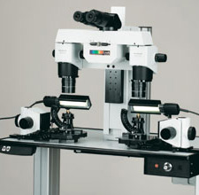
Comparison microscope
Before the use of comparison microscopy, forensic firearms examinations typically involved
- the sequential examination of fired components using a single compound microscope,
- large format photography of microscopic details for each component through a compound microscope,
- side-by-side comparison of the photographic results,
- preparation of exhibits based on the photographs.
Problems associated with this approach included these:
- The evidence items were examined in sequence, not simultaneously.
- The evidence items could only be simultaneously compared using photographic prints.
- The photographs taken were two-dimensional representations of three-dimensional objects.
Early in 1925, Colonel Calvin Goddard (generally acknowledged as the father of firearms identification in the United States) and his associate, Phillip O. Gravelle, (microscopist, tool designer, and photographer) adapted the existing compound microscope to accommodate simultaneous microscopic forensic bullet comparisons. They achieved this by using an optical bridge to join together the stages of two compound microscopes.
This basic monocular instrument has evolved considerably over the past eighty years into the sophisticated instruments used today.
Some of the features:
- Binocular viewing
- Rotating nosepieces with a variety of objective lenses
- A choice of illumination systems, e.g., variable fiber optic, LED, and high-intensity fluorescent light sources
- Push-button focusing
- Image capture systems with file export and printing capabilities
- Specialized specimen mounts
- Motorized control of the x, y, and z axis settings, which can be saved for reproducibility
- Monitors to facilitate examinations, training, and image capture
- Optical and digital capability for image superimposition or conventional side-by- side comparisons
- Digital reference marks to easily return to areas of interest